Zeiss AxioObservor Z1 Epifluorescence Microscope
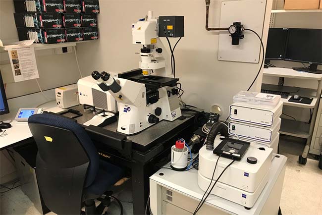
• The LCBG Core Facility houses a Carl Zeiss LSM 780 laser scanning confocal mounted on a motorized AxioObserver Z1 inverted microscope. The confocal microscope is equipped with two PMTs and one 32 channel GaAsP spectral detector. It has 7 laser lines (405nm, 458nm, 488nm, 514nm, 543/561nm, 594nm & 633nm), and an automated scanning stage.
• The microscope is equipped with a variety of objectives including an EC Plan Neoflaur 10x/0.3 DICI, Plan Apochromat 20x/0.8 DICII, a Plan Apochromat 40x/1.4 DIC, Plan Apochromat 63x/1.4 Oil DIC, and Plan Neofluar 100x/1.3 Oil DIC.
• The microscope is equipped with a stage top incubator system to control temperature, humidity, and CO2 levels for live cell imaging.
• The major uses of a confocal microscope include 3D reconstruction and animation of photosections generated from thick specimens, studying dynamics of molecular mobility by Fluorescence Recovery after Photobleaching (FRAP) and Fluorescence Loss in Photobleach (FLIP), and examining molecular interactions using Fluorescence Resonance Energy Transfer (FRET).
• Images are acquired and analyzed using Carl Zeiss Zen Black software.
Zeiss AxioObservor Z1 Epifluorescence Microscope
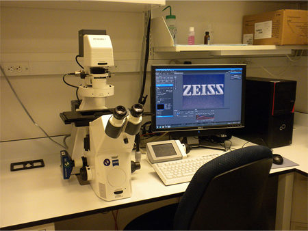
• The LCBG Microscopy Core Facility has two Carl Zeiss AxioObservor Z1 inverted microscopes used for phase contrast, DIC, and fluorescent imaging.
• The microscopes are equipped with either an AxioCam MRm monochrome camera or an AxioCam 506 color camera.The available objectives include a Fluar 5x/0.25, an N Achroplan 10x/0.25 Ph1, an LD Plan Neofluar 20x/0.4 Ph2, an LD Plan Neofluar 40x/0.6 Corr Ph2, an EC Plan Neoflaur 40x/1.3 Oil DIC, and a Plan Apochromat 63x/1.4 Oil DIC.
• Filter cubes installed on the microscopes allow for up to four color imaging using DAPI, GFP, Cy3, and Cy5.
• Stage inserts allow imaging of live or fixed cells in multiwell plates, or dishes up to 10cm.
• Images are acquired and analyzed using Carl Zeiss Zen Blue software.
Nikon Eclipse Ti2-E Inverted Microscope
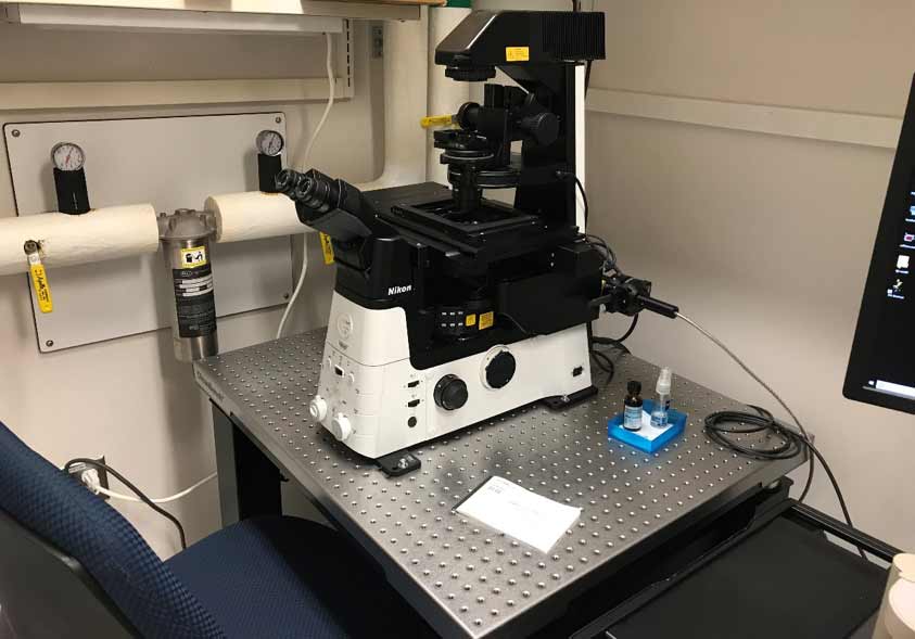
• The LCBG Microscopey Core houses a Nikon Eclipse Ti2-E inverted microscope acquired by the facility in 2018.
• The microscope is optimized for single molecule RNAFISH imaging and is equipped with a Photometrics Prime BSI sCMOS camera and a Lumencore SOLA SE 365 FISH light engine.
• The microscope has a motorized scanning stage with Z piezo insert for fast Z and multi position time lapse imaging.
• The available objectives are all plan Apochromat, and include a 10x/0.45 DIC, 20x/0.75 DIC, 40x/1.3 Oil DIC, , 60x/1.4 Oil DIC, and a 100x/1.45 Oil DIC objective.
• Filter cubes installed on the microscope are narrow band optimized for FISH imaging using DAPI, FITC, Gold, Red, and Cy5.
• Images are acquired and analyzed using Nikon Elements software.
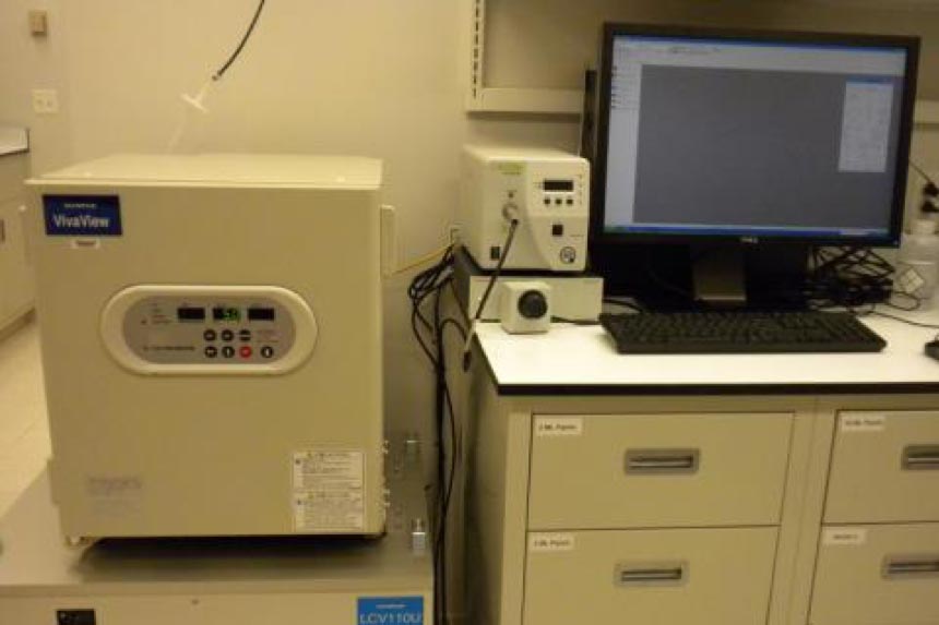
Olympus VivaView FL Incubator Fluorescence Microscope
• Provides a fully integrated and motorized inverted microscope to allow high quality, long-term time-lapse imaging in a constant and optimized environment, including hypoxia.
• Equipped with a UPLSAPO40x objective with intermediate 0.5x/1x/2x magnification.
• Multiple locations in up to 8 samples can be imaged simultaneously with fluorescence (DAPI, GFP, DsRED) or differential interference contrast (DIC).
• Multi-gas capability allows control of O2 levels as well as CO2.
• Time lapse images are acquired and analyzed using Olympus Metamorph software.
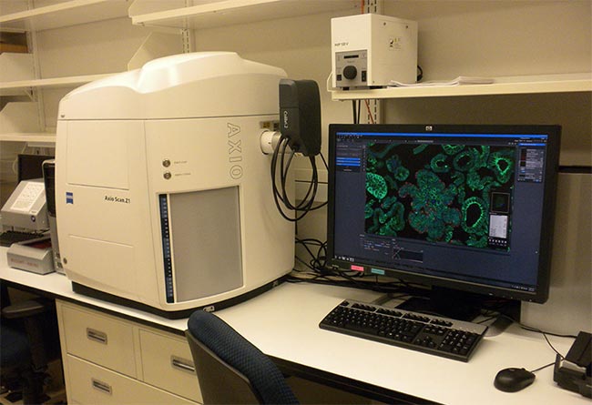
Zeiss AxioScan Z1 Slide Scanning Microscope
• The LCBG Microscopy Core Facility has a Carl Zeiss AxioScan Z1 fully automated slide scanning microscope able to create brightfield and fluorescent virtual slides in high quality and at speed.
• The system is equipped with a Colibri 7 LED source with 385nm, 430nm, 475nm, 511nm, 555nm, 590nm and 630nm LEDs
• The microscope has a number filter cubes allowing the use of dyes including DAPI, FITC, TRITC, Cy5, CFP, YFP and mCherry.
• Available objectives includes a Fluar 5X/0.25, a Plan Apochromat 10x/0.3, a Plan Apochromat 20x/0.3, and a Plan Apochromat 40x/0.95 Corr.
• Up to 100 slides can be digitized at one time, and images are acquired and analyzed using Carl Zeiss Zen 2.3 software.
