Software

LAS
Leica Application Suite X (LAS X) is the one software platform for all Leica microscopes: It integrates confocal, wide field, stereo, super-resolution, and light-sheet instruments from Leica Microsystems.
MetaMorph
The MetaMorph® Microscopy Automation and Image Analysis Software automates acquisition, device control, and image analysis.
Elements
Nikon’s universal software platform, NIS-Elements combines powerful image acquisition, analysis, visualization and data sharing tools.

Vision4D
Arivis Vision4D is a modular software for working with multi-channel 2D, 3D and 4D images of almost unlimited size independent of available RAM.
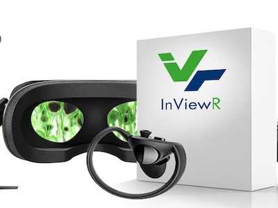
Arivis InViewR
arivis InViewR sets new standards for analyzing Life Science research images. Freed from being tethered to a mouse like on a desktop computer and with depth perception equivalent to the real word.
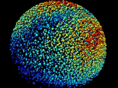
Imaris
The world’s leading Interactive Microscopy Image Analysis software company, actively shaping the way microscopic images are processed through constant innovation and a clear focus on 3D and 4D imaging.
Matlab
MATLAB® combines a desktop environment tuned for iterative analysis and design processes with a programming language that expresses matrix and array mathematics directly.

Huygens Deconvolution
Deconvolution is a mathematical operation used in Image Restoration to recover an image that is degraded by a physical process which can be described by the opposite operation, a convolution.
Techniques
Immunofluorescence
Technique used to assess both the localization and endogenous expression levels of their favorite proteins.
Live cell imaging (particularly T cell activation assays)
Live cell imaging is the study of living cells using time-lapse microscopy. It is used by scientists to obtain a better understanding of biological function through the study of cellular dynamics.
Photobleaching and Photo-activation
Photobleaching
Involves bleaching a region of interest in a sample with intense light, and then monitoring the fluorescence over time. This technique can be used to study the mobility of molecules, such as proteins, in cells. Photobleaching can occur during imaging after photo-activation, so the power and duration of the photo-activation must be optimized to minimize photobleaching
Photoactivation
Involves using a brief, high-intensity light pulse to convert a photoactivatable fluorescent protein (PAFP) into a different fluorescent state. The PAFPs can be tagged to a host protein, and then followed over time as they re-equilibrate in the cell. Photo-activation is a complementary strategy to Fluorescence Recovery After Photobleaching (FRAP).
FRET
FRET is a technique to detect protein interaction and the formation of complexes between different proteins tagged with fluorescence markers. It also can be used to detect enzymatic activity by special reporters (FRET biosensors).
TIRF
A total internal reflection fluorescence microscope (TIRFM) is a type of microscope with which a thin region of a specimen, usually less than 200 nanometers can be observed.
SMLM
The multiple imaging modes afforded by widefield, confocal, and multiphoton fluorescence microscopies permit noninvasive, temporally resolved imaging of fixed and living cells and tissues with a high level of biochemical specificity.