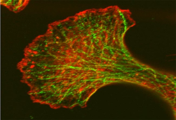
Confocal Microscope Image of Fixed Mouse Embryonic Fibroblast (MEF) Cells
Actin proteins (stained red) and Tubulin proteins (stained green) are involved in a number of cellular process such as cell motility, cell division and maintenance of cell shape. Both proteins form dimers which form micofilaments (actin) and microtubules (tubulin). They are potential targets for cancer therapy. Cells prepared and imaged by Dr. Luanna Scheffer, laboratory of Dr. Jairaj Acharya, CCR, FNLCR
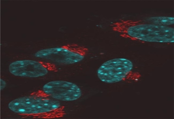
Confocal Microscope Image of Fixed Mouse Embryonic Fibroblast (MEF) Cells
The golgi complex (stained red) and the nucleus (stained cyan). The golgi complex modifies, sorts, and packages proteins for cell secretion or use inside the cell. It also plays roles in lipid transport, lysosome creation and proteoglycan synthesis. Samples prepared and imaged by Dr. Luana Scheffer, laboratory o f Dr. Jairaj Acharya, CCR, FNLCR.
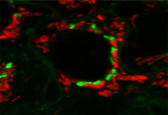
Confocal Microscope Image of Live Mouse Embryonic Fibroblast (MEF) Cells
The golgi apparatus (stained green) modifies, sorts and packages proteins for cell secretion or use inside the cell. It also plays roles in lipid transport, lysosome creation and proteoglycan synthesis. Mitochondria (stained red) converts energy into forms that are usable by the cell. It plays roles in cellular respiration as well as cell division, growth and death. Samples prepared and imaged by Dr. Luana Scheffer, laboratory o f Dr. Jairaj Acharya, CCR, FNLCR.
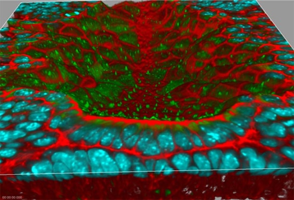
3-D Rendering of the Node of a Mouse Embryo
The red staining is F-actin, the green is antibody labeling of acetylated tubulin to visualize cilia, and the cyan is cell nuclei. The sample preparation and labeling were performed in the laboratory of Dr. Terry P. Yamaguchi, Cancer and Developmental Biology Laboratory, Center for Cancer Research, National Cancer Institute. Image acquisition and 3-D rendering were performed in the laboratory of Dr. Stephen Lockett, Advanced Technology Program FNLCR/SAIC-Frederick.
Confocal Microscope Image of Bovine Pulmonary ArteryEndothelial (BPAE) Cells
(BPAE) cells stained with a combination of fluorescent dyes. Mitochondria were labeled with red-fluorescent MitoTracker Red CMXRos, F-actin was stained using green-fluorescent Alexa Fluor 488 phalloidin, and blue-fluorescent DAPI was used to label the nuclei. Endothelial cells line the blood vessels and provide a barrier between circulating blood and the rest of the vessel wall. Among the processes they are involved in are angiogenesis, vasculogenesis, vasoregulation and blood cell trafficking. Cells are Invitrogen’s FluoCells prepared slide #1 (F36924).
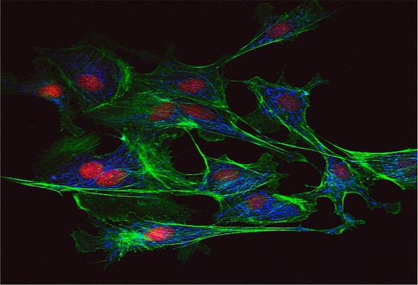
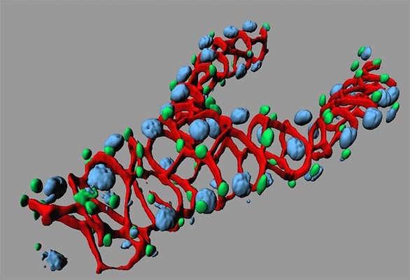
Image of Drosophila Kidney RC Cells
Red channel is antibody-labeled Scrib in RC cells (one of the tumor suppressors in Drosophila) antibody-labeled in Drosophila kidney RC cells; Green is Stat-GFP, which labels the kidney stem cells (RNSC) and transit cells (RB); Blue is DAPI. Sample prepared and images by Dr. Xiankun Zeng, laboratory of Dr. Steven Hou, CCR, FNL and rendered with IMARIS.
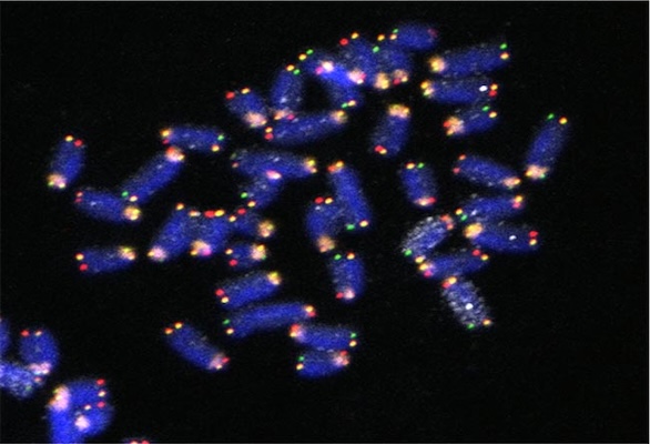
Five-color FISH Imaging in Mouse Embryonic Stem Cells to study Strand Segregation in Chromosome 11 through Mitosis.
DNA (blue), Centromere (orange), Chromosome 11 (white), G repeat Telomeres (green), C repeat Telomere (red). Metaphase spread was prepared so that one sister chromatid is single stranded. Sample prepared and imaged by Dr. Stephen Sauer, laboratory of Dr. Amar Klar, CCR, NCI-Frederick.
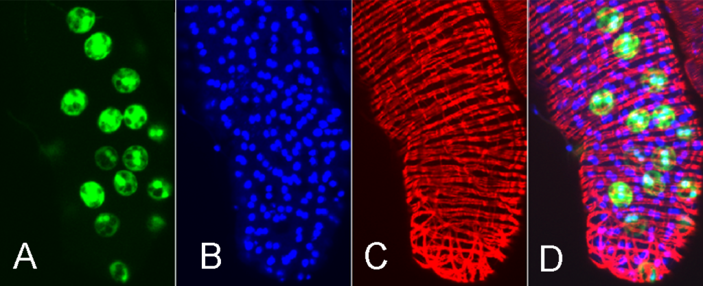
Images of Drosophila Prostate
A) Escargot-GFP labeling a subset of cells. B) DAPI nuclear staining. C) Red: phalloidin labeling of F actin. D) Overlay. Sample prepared and imaged by Dr. Shree Ram Singh, Laboratory of Dr. Steven Hou of CCR, NCI-Frederick (unpublished)