Our Team

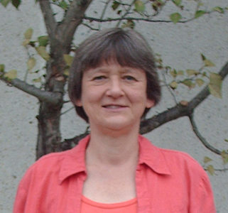
Tatiana S. Karpova Ph.D.
Core Head
karpovat@nih.gov
Building 41, Room C615
240-760-6637
Education
• St. Petersburg University – B.S./M.S. (Genetics) – 1973-1979
• St. Petersburg University – Ph.D. (Genetics) – 1979-1979
Honors
• FEBS Fellowship (Vrije Univ., Amsterdam, Holland) – 1991
• American Heart Association Fellowship (Washington Univ., St. Louis, MO) – 1993-1995
• NIH/NCI Group Award of Merit (Bethesda, MD) – 2005
Research Experience
• 1979-1984: Graduate student/Biologist, Department of Genetics, St. Petersburg University, Russia. Studied the transfer of cytoplasmic factors by cytoduction in yeast with S. G. Inge-Vechtomov, St. Petersburg University, Russia. Applied genetic methods (micromanipulation and tetrad analysis, mutagenesis, cytoduction).
• 1984-1991: Assistant Professor; Department of Genetics, St. Petersburg University, Russia. Studied the genetics and molecular biology of chromosomal stability in yeast, St. Petersburg University, Russia. Applied genetic methods (micromanipulation and tetrad analysis, mutagenesis) and molecular biology methods (plasmid and strain construction).
• 1992-1999: Post doctorate in the lab of J. Cooper; Washington University Medical School, St. Louis, MO, USA. Studied the dynamics and genetic control of depolarization of actin cytoskeleton. Used methods of molecular biology (sequencing, Northern, Western analysis, plasmid construction, fluorescence microscopy in live cells, deconvolution, immunofluorescence of yeast, mutagenesis). First publication on the application of GFP to studies of the yeast actin cytoskeleton in collaboration with J. Waddle.
• 1999-present: Manager of the Optical Microscopy Core (OMC)/ Biologist; NCI/NIH, Bethesda, MD, USA. Studied the regulation of transcription in yeast, using CUP1 locus as a model in the lab of J. G. McNally. Used methods of deconvolution, live imaging with fluorescent markers, single molecule FISH and FRAP in research. Managed the Optical Microscopy Facility and applied FRET, FRAP, fluorescence imaging and quantification and cell tracking techniques.
Professional Activities
• FAES lecturer in FRET and GFP and live imaging microscopy
• Member of ORSAC (Office of Research Support Advisory Committee)• St. Petersburg University – Ph.D. (Genetics) – 1979-1979
Invited Lectures
• Gordon Conference, Plant and Fungal Cytoskeleton, Andover, NH, 1995
• Light Microscopy Interest Group, NIH, Bethesda, MD, 2003
• Chromatin Interest Group, NIH, Bethesda, MD, 2008
• Transcription Interest Group, NIH, Bethesda, MD, 2008
• Faculty/Post-doctoral Seminar Series, University of South Dakota Sanford School of Medicine, Vermillion, SD, 2008
• Eppley Seminar Series Speaker, Eppley Institute for Cancer Research, University of Nebraska Medical Center , Omaha, NE, 2008
• Light Microscopy Interest Group, NIH, Bethesda, MD, 2013• St. Petersburg University – Ph.D. (Genetics) – 1979-1979
Selected Publications
1. FRAP: Tatiana S. Karpova, Teresa Y. Chen, Brian L. Sprague, James G. McNally. Dynamic interactions of a transcription factor with DNA are accelerated by a chromatin remodeler. EMBO Reports. 5: 1064-1070, 2004• St. Petersburg University – Ph.D. (Genetics) – 1979-1979
2. Deconvolution: Arnaoutov A, Azuma Y, Ribbeck K, Joseph J, Boyarchuk Y, Karpova T, McNally J, Dasso M. Crm1 is a mitotic effector of Ran-GTP in somatic cells. Nature Cell Biol 7: 626-632, 2005
3. FRET: Karpova, T.S. and McNally, J.G. Detecting protein-protein interactions with CFP-YFP FRET by acceptor photobleaching. Curr. Prot. Cytom ; Chapter 12: Unit 2.7 (12.7.1-12.7.11), 2006
4. FRET: Koga F., Xu W, Karpova TS, McNally JG, Baron R, Neckers L. Hsp90 inhibition transiently activates Src kinase and promotes Src-dependent Akt and Erk activation. Proc Natl Acad Sci U S A. 103(30):11318-22, 2006.
5. FRAP and Live Imaging: Tatiana S. Karpova, Min J. Kim, Corentin Spriet, Kip Nalley, Tim Stasevich, Zoulika Kherrouche, Laurent Heliot, and James G. McNally Concurrent Fast and Slow Cycling of a Transcriptional Activator at an Endogenous Promoter. Science 319: 466-469, 2008
6. FRAP: Sprouse RO, Karpova T, Mueller F, Dasgupta A, McNally JG, Auble DT. Regulation of TATA binding protein dynamics in living yeast cells.PNAS, 105:13304-13308, 2008
7. Immunofluorescence: Mark Rochman, Yuri Postnikov, Sarah Correll, Cedric Malicet, Stephen Wincovitch, Tatiana S. Karpova, James G. McNally, Xiaolin Wu, Nina A. Bubunenko, Sergei Grigoryev, Michael Bustin. The Interaction Of NSBP1 With Nucleosomes In Euchromatin Counteracts Linker Histone-Mediated Chromatin Compaction And Modulates transcription. Mol Cell 35: 642-656, 2009
8. FRAP: Dey A., Nishiyama, A., Karpova, T., McNally, J., Ozato, K. Brd4 marks select genes on mitotic chromatin and directs postmitotic transcription. Mol Biol Cell. 20: 4899-4909, 2009
9. FRET biosensors: Komiya, T., Coxon, A., Park,Y., Chen, W. D., Zajac-Kaye, M., Meltzer, P., Karpova, T., Kaye, F. J. Enhanced activity of the CREB co-activator Crtc1 in LKB1 null lung cancer. Oncogene, 29: 1672-80, 2010
10. FRET: Heyerdahl, S., Rosenberg, J., Jamtgaard, L., Rishi, V., Varticovski, L., Akah, K., Scudiero, D., Shoemaker, R. H., Karpova, T., Day, R. N., McNally, J. G., Vinson, C. The arylstibonic acid compound NSC13746 disrupts B-ZIP binding to DNA in living cells. Eur J Cell Biol 89:564-573, 2010. PMID: 20362353
11. Immunofluorescence: Stuelten, C. H., Busch, J., Tang, B., Flanders, K., Oshima, A., Sutton, E., Karpova, T.S., Roberts, A.B., Wakefield, L. M., Niederhuber, J. E. Transient tumor-fibroblast interactions increase tumor cell malignancy by a TGF-beta mediated mechanism in a mouse xenograft model of breast cancer. PLoS One, 5: e9832, 2010
12. Image Quantification: Gordon S. N., Cecchinato V., Andresen V., Heraud J-M., Hryniewicz A., Parks R.W., Venzon D., Chung H., Karpova T., McNally J., Silvera P., Reimann K. A., Matsui H, Kanehara T., Shinmura Y., Yokote H., Franchini G. Smallpox Vaccine safety is dependent on T cells and not B cells. J Infectious diseases (JID), 203: 1043-1053, 2011
13. Live Cell Imaging: Tatiana A. Chernova, Andrey V. Romanyuk, Tatiana S. Karpova, Nela Moffatt, John R. Shanks, Moiez Ali, Andrew O’Dell, James G. McNally, Susan W. Liebman, Yury O. Chernoff, Keith D. Wilkinson. Prion Induction by the Short-lived Stress Induced Protein Lsb2 Is Regulated by Ubiquitination and Association with the Actin Cytoskeleton. Mol Cell 43: 242-252, 2011.
14. FCS, TICS methods: Mazza D, Stasevich TJ, Karpova TS, McNally JG. Monitoring dynamic binding of chromatin proteins in vivo by fluorescence correlation spectroscopy and temporal image correlation spectroscopyMethods Mol Biol. 833:177-200, 2012.
15. FRAP methods: Mueller F, Karpova TS, Mazza D., McNally JG. Monitoring dynamic binding of chromatin proteins in vivo by fluorescence recovery after photobleaching. Methods Mol Biol. 833:153-176, 2012.
16. Live Cell Imaging and Tracking: Hurley A., Smith M., Karpova T, ……McNally J, ….Catalfamo M. Enhanced effector function of CD8(+) T cells from healthy controls and HIV-infected patients occurs through thrombin activation of protease-activated receptor 1. J Infect Dis. 207: 638-650, 2013.
17. SMT: Ball, D.A., Mehta, G. D., Salomon-Kent, R., Mazza, D., Morisaki, T., Mueller, F., McNally, J.G., Karpova, T. S.Single Molecule Tracking Of Ace1p In Saccharomyces cerevisiae Defines A Characteristic Residence Time For Non-specific Interactions Of Transcription Factors With Chromatin. (2016) Nucleic Acids Res 44:e160.
18. SMT: Presman, D., Ball, D., Paakinaho, V., Grimm, J., Lavis, L., Karpova, T., Hager, G.: Quantifying transcription factor dynamics at the single-molecule level in live cells. (2017) Methods 123: 76-88
19. FRET and biosensors:.Kannan R., Song JK, Karpova T, Clarke A, Shivalkar M, Wang B, Kotlyanskaya L, Kuzina I, Gu Q, Giniger E. The Abl pathway bifurcates to balance Enabled and Rac signaling in axon patterning in Drosophila. (2017) Development 144: 487-498
20. SMT: Paakinaho, V., Presman DM, Ball D, Schiltz, RL, Levitt, P, Johnson, T, Mazza D., Morisaki T, Karpova TS, Hager GL. Single-Molecule analysis of steroid receptor and cofactor actin in living cells. (2017). Nature Communications 8: 15896.
21. Quantification of cells in tissues: Rahman, M.A., McKinnon, K.M., Karpova T.S., Ball D.A., Venzon, D. J. Fan, W., Kang, G, Li, Q., Robert-Guroff, M. Associations of Simian Immunodeficiency Virus (SIV)-Specific follicular CD8 T cells with Other Follicular T cells suggest complex contributions to SIV Viremia control.(2018) J. Immunol. 200: 2714-2726.
22. SMT: Serebryannyy, L. A., Ball, D. A., Karpova T. S., Misteli, T. Single molecule analysis of lamin dynamics (2018) Methods, 157:56-65.
23. SMT and smFISH: Mehta G.D., Ball D. A., Eriksson, P. R., Chereji, R. V., Clark D. J., McNally J. G., Karpova, T. S. Single-Molecule analysis reveals linked cycles of RSC chromatin remodeling and Ace1p transcription factor binding in yeast (2018) Molecular Cell 72: 875-887.

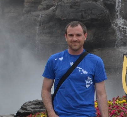
David A. Ball Ph.D.
Core Biologist
balla@nih.gov
Building 41, Room B114D
240-760-6577
Education
• University at Albany (SUNY) – B.S. (Physics) – 1996-2000
• University of Tennessee Space Institute – M.S./Ph.D. (Physics) – 2000-2006
Honors
• Sigma Pi Sigma Physics Honor Society
Research Experience
2000-2006: Graduate Research Assistant, University of Tennessee Space Institute
Training in general physics with an emphasis on optics. Research focused on the development of instrumentation and data-reduction methods to use the sensitive detection of fluorescence by confocal microscopy as a means of monitoring molecular dynamics.
• Constructed custom microscope for Single-Molecule Detection and Imaging by Laser-Induced Fluorescence using both epi-illumination and prism-based total internal reflection (TIRF).
• Aided in development of data collection and analysis software for Fluorescence Correlation Spectroscopy (FCS).
• Developed software to a) analyze temporal brightness fluctuations (blinking) of single fluorescent molecules, and b) deconvolve scattered light signal from time-correlated single-photon counting (TCSPC) measurements to extract fluorescence lifetime.
2007-2010: Postdoctoral Associate, Virginia Bioinformatics Institute
Live cell imaging to measure expression of proteins regulating the cell cycle in Saccharomyces cerevisiae, and the characterization of synthetic biology constructs in Escherichia coli.
• Developed image processing algorithm to automatically identify cells in phase-contrast microscope images, and track changes in their mean and localized fluorescence over time.
• Trained and mentored undergraduate and high-school students in the use and analysis of microscopy, flow cytometry and bulk fluorescence measurements for investigations of gene expression.
2011-2014: Senior Research Associate, Virginia Bioinformatics Institute
Live- and fixed-cell imaging to study cell cycle regulation in Saccharomyces cerevisiae, with added responsibilities of laboratory management and software development.
• Developed algorithms to automatically count in hundreds of individual yeast cells the number of mRNA molecules from cell cycle regulating genes tagged by single-molecule fluorescence in situ hybridization (FISH), and detected with fluorescence microscopy.
• Led the development of GenoSIGHT, an adaptive microscope control system written in MATLAB to automate the characterization of gene expression in live cells.
• Created models of gene networks using both COPASI and MATLAB’s SimBiology toolbox
• Directly supervised a team of four undergraduate students as part of the 2011 International Genetically Engineered Machines (iGEM) competition. The goal of the students’ project was to characterize the maturation kinetics of multiple fluorescent proteins in both yeast and bacteria.
2014-present: Staff Scientist, OMC .
Develops methods of quantification of Single Molecule Tracking, smFIS data modeling. Supports SMT and cell segmentation in tissues. Builds and maintains custom microscopes.
Professional Activities at the NCI/NIH
Dr. Ball provides expert advice and support to Core customers on data collection, data analysis and quantification including but not limited to quantitative analysis of high-resolution statistical mapping of the super-resolution images, FRAP, FCS, RICS, Single molecule tracking, Number and Brightness analysis, and Single Molecule FISH quantification. Dr. Ball is an expert in assembling prototype microscopes that are not yet available commercially and that utilize new optical principles and technical design. Currently Dr. Ball develops instrumentation for the Single Molecule Tracking/Super-resolution microscope and TILT lattice
Invited Lectures
• Kongju National University, Department of Chemistry, Kongju, South Korea, 2006
Selected Publications
1. Feedback-controlled image acquisition: “Adaptive imaging cytometry to estimate parameters of gene networks models in systems and synthetic biology,” D.A. Ball, M.W. Lux, N.R. Adames, and J. Peccoud, PLoS ONE. 9 (9): e107087 (2014)• University of Tennessee Space Institute – M.S./Ph.D. (Physics) – 2000-2006
2. FISH: “Measurement and modeling of transcriptional noise in the cell cycle regulatory network,” D.A. Ball, N.R. Adames, N. Reischmann, D. Barik, C.T. Franck, J.J. Tyson, and J. Peccoud, Cell Cycle, 12 (19), 3203 (2013).
3. Live Imaging: “Oscillatory dynamics of cell cycle proteins in single yeast cells analyzed by imaging cytometry,” D.A. Ball, J. Marchand, M. Poulet, W.T. Baumann, K.C. Chen, J.J. Tyson, and J. Peccoud, PLoS ONE, 6 (10): e26272 (2011).
4. Live imaging and Modeling: Stochastic exit from mitosis in budding yeast: Model predictions and experimental observations. D.A. Ball, T.-H. Ahn, P. Wang, K.C. Chen, Y. Cao, J.J. Tyson, J. Peccoud and W.T. Baumann, Cell Cycle, 10 (6), 999 (2011).
5. Feedback-controlled image acquisition: Cyto•IQ: An adaptive cytometer for extracting the noisy dynamics of molecular interactions in live cells. D.A. Ball, S.E. Moody, and J. Peccoud, in Imaging, Manipulation, and Analysis of Biomolecules, Cells, and Tissues VII, D.L Farkas, D.V. Nicolau, and R.C. Leif, eds., Proc. SPIE, 7568, 75681D (2010).
6. Single Molecules: “Single-Molecule Detection with axial flow near micron-sized capillaries,” D. A. Ball, G. Shen, and L. M. Davis, Appl. Opt., 46, 1157-1164 (2007)
7. FCS: Data reduction methods for application of fluorescence correlation spectroscopy to pharmaceutical drug discovery. L.M. Davis, P.E. Williams, D.A. Ball, E.D. Matayoshi, and K.M. Swift, Curr. Pharm. Biotech. 4, 451-462 (2003).
8. FCS: Dealing with reduced data acquisition times in fluorescence correlation spectroscopy for HTS applications. L. M. Davis, D. A. Ball, P. E. Williams, E. D. Matayoshi, and K. M. Swift, in Microarrays and Combinatorial Technologies for Biomedical Applications, D. V. Nicolau and R. Raghavachari, eds., Proc. SPIE, 4966, 117-128 (2003)
9. Single Molecules: Imaging of single-chromophore molecules in aqueous solution near a fused-silica interface. L.M. Davis, W.C. Parker, D.A. Ball, J.G.K. Williams, G.R. Bashford, P. Sheaff, R. Eckles, D.T. Lamb, and L.R. Middendorf, Proc. SPIE, 4262, pp. 301-311 (2001)
10. SMT: Ball, D.A., Mehta, G. D., Salomon-Kent, R., Mazza, D., Morisaki, T., Mueller, F., McNally, J.G., Karpova, T. S. Single Molecule Tracking Of Ace1p In Saccharomyces cerevisiae Defines A Characteristic Residence Time For Non-specific Interactions Of Transcription Factors With Chromatin. (2016) Nucleic Acids Res 44:e160.
11. SMT: Presman, D., Ball, D., Paakinaho, V., Grimm, J., Lavis, L., Karpova, T., Hager, G.: Quantifying transcription factor dynamics at the single-molecule level in live cells. (2017) Methods 123: 76-88 PMID: 28315485
12. SMT: Paakinaho, V., Presman DM, Ball D, Schiltz, RL, Levitt, P, Johnson, T, Mazza D., Morisaki T, Karpova TS, Hager GL. Single-Molecule analysis of steroid receptor and cofactor actin in living cells. (2017). Nature Communications 8: 15896.
13. Quantification of cells in tissues: Rahman, M.A., McKinnon, K.M., Karpova T.S., Ball D.A., Venzon, D. J. Fan, W., Kang, G, Li, Q., Robert-Guroff, M. Associations of Simian Immunodeficiency Virus (SIV)-Specific follicular CD8 T cells with Other Follicular T cells suggest complex contributions to SIV Viremia control.(2018) J. Immunol. 200: 2714-2726.
14. SMT: Serebryannyy, L. A., Ball, D. A., Karpova T. S., Misteli, T. Single molecule analysis of lamin dynamics (2018) Methods, 157:56-65
15. SMT and smFISH: Mehta G.D., Ball D. A., Eriksson, P. R., Chereji, R. V., Clark D. J., McNally J. G., Karpova, T. S. Single-Molecule analysis reveals linked cycles of RSC chromatin remodeling and Ace1p transcription factor binding in yeast (2018) Molecular Cell 72: 875-887

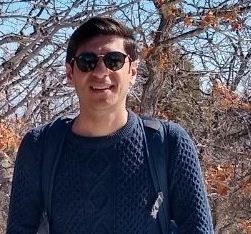
Mohamadreza Fazel, Ph.D.
Core Biologist
mohamadreza.fazel@nih.gov
Building 41, Room B114D
240-858-7546
Education
• Shahrekord University – B.S. (Physics) – 2006-2010
• Isfahan University of Technology — M.S. (Physics) – 2010-2013
• University of New Mexico – M.S. (Optical Engineering) – 2014-2016
• University of New Mexico – Ph.D. (Physics) – 2016-2020
Honors
• Honor student at Shahrekord University for maintaining a high GPA throughout the program, 2010
• Graduate Studies Excellence Assistantship (GSEA), University of New Mexico, 2017
• S-CAP trave; award, University of New Mexico, 2019
Research Experience
2014-2016 graduate teaching assistant, University of New Mexico
• Teaching multiple courses and introductory physics labs
2016-2020 graduate research assistant, University of New Mexico
• Training in a biophysics lab where I worked on the analysis of super-resolution single molecule localization microscopy data.
• Developed a multi-emitter localization method to pinpoint emitters within densely regions with overlapping PSFs and structured backgrounds in SMLM data
• Developed a Bayesian method to achieve nanometer localization precisions using SMLM data
• Involved in developing a comprehensive data processing and post-processing pipeline for SMLM data.
• Involved in the development of a drift correction tool as a postprocessing of SMLM data.
2020-2024 postdoctoral research scholar, Arizona State University
• Developing tools for fluorescent lifetime imaging microscopy to achieve high spatial and lifetime resolutions while learning the number of species present within the data
• Involved in developing a comprehensive methodological framework for FRET data analysis capable of inferring the number of conformational states.
• Developing a 3D tracking frameworks using aberrated PSFs.
• Developing a computational framework for spectral-FLIM data analysis capable of learning dealing with many species with overlapping spectra.
2024-presnt staff scientists, National Cancer Institute, Laboratory of Receptor Biology and Gene Expression
• Developing computational tools for the processing and post-processing of MINFLUX and DNA-PAINT data.
Professional Activities at the NCI/NIH
As a staff scientist at the optical microscopy core (OMC) in the Laboratory of Receptor Biology and Gene Expression. He has a strong interdisciplinary background in applying cutting-edge statistical machine learning methods to address problems in biophysics, fluorescence microscopy and optics. At OMC, his research is mainly focused on pattern recognition in data from advanced nanoscopy methods, such as MINFLUX and DNA-PAINT.
Invited Lectures
Invited Zoom Talk—LFD Workshop, University of California, Irvine, California, USA, November 2023.
Popular Science Talk—Science On Tap, Tempe, AZ, USA, October 2023.
Invited Zoom Talk—Physics Seminar, University of Calgary, Alberta, Canada, January 2022.
Invited Zoom Talk–Physics Colloquium, Isfahan University of Technology, Isfahan, Iran, October 2021.
Selected Publications
1. STORM: Hamidreza Heydarian, Florian Schueder, Maximilian T. Strauss, Ben van Werkhoven, Mohamadreza Fazel, Keith A. Lidke, Ralf Jungmann, Sjoerd Stallinga, Bernd Rieger, ‘Template-free 2D particle fusion in localization microscopy’. Nature Methods 09/2018; 15(10). DOI: 10.1038/s41592-018-0136-6.
2. STORM: Mohamadreza Fazel, Michael J. Wester, Hanieh Mazloom-Farsibaf, Marjolein B. M. Meddens, Alexandra Eklund, Thomas Schlichthaerle, Florian Schueder, Ralf Jungmann, Keith A. Lidke, ‘Bayesian multiple emitter fitting using reversible jump Markov chain Monte Carlo’. Scientific Reports 9, 13791 (2019). DOI: 10.1038/s41598-019-50232-x.
3. STORM: Hanieh Mazloom-Farsibaf, Farzin Farzam, Mohamadreza Fazel, Michael J. Wester, Marjolein Meddens, Keith A. Lidke, ‘Comparing lifeact and phalloidin for super-resolution imaging of actin in fixed cells’, PLoS One 16(1), e0246138 (2021). DOI:
4. STORM: Michael J. Wester, Sandeep Pallikkuth, Hanieh Mazloom-Farsibaf, Mohamadreza Fazel, David Schodt and Keith A. Lidke, ‘Robust, fiducial free drift correction for super-resolution imaging’, Scientific Reports 11, 23672 (2021). DOI: 10.1038/s41598-021-02850-7.
5. STORM: Mohamadreza Fazel, Michael J. Wester, ‘Analysis of super-resolution single molecule localization microscopy data: A tutorial’, AIP Advances 12(1):010701 (2022). DOI: 10.1063/5.0069349. (This work was featured as the most downloaded review paper from the journal in 2022).
6. FLIM: Mohamadreza Fazel, Sina Jazani, Lorenzo Scipioni, Alexander Vallmitjana, Enrico Gratton, Michelle A. Digman, and Steve Pressé, ‘High resolution fluorescence lifetime maps from minimal photon counts’, ACS Photonics (2022). DOI: 10.1021/acsphotonics.1c01936.
7. STORM: William K. Kanagy, Cédric Cleyrat, Mohamadreza Fazel, Shayna R. Lucero, Marcel P. Bruchez, Keith A. Lidke, Bridget S. Wilson, and Diane S. Lidke, ‘Docking of Syk to FcεRI is enhanced by Lyn but limited in duration by SHIP1’, Molecular Biology of the Cell (2022). DOI: 10.1091/mbc.e21-12-0603.
8. STORM: Mohamadreza Fazel, Michael J. Wester, David J. Schodt, Sebastian Restrepo Cruz, Sebastian Strauss, Florian Schueder, Thomas Schlichthaerle, Jennifer M. Gillette, Diane S. Lidke, Bernd Rieger, Ralf Jungmann, Keith A. Lidke, ‘High-Precision Estimation of Emitter Positions using Bayesian Grouping of Localizations’, Nature Communications 13 (1), 7152 (2022). DOI: 10.1038/s41467-022-34894-2.
9. FRET: Ayush Saurabh, Matthew Safar, Ioannis Sgouralis, Mohamadreza Fazel, Steve Pressé, “Single photon smFRET. I. theory and conceptual basis”, Biophysical Reports 3 (1), 100089 (2022). DOI: 10.1101/2022.07.20.500887. (This work was featured on the journal’s cover and was highlighted in the Biophysical Society website as cornerstone research).
10. FRET: Ayush Saurabh, Matthew Safar, Mohamadreza Fazel, Ioannis Sgouralis, Steve Pressé, “Single photons smFRET. II. Application to continuous illumination”, Biophysical Reports 3 (1), 100087 (2022). DOI: 10.1101/2022.07.20.500888. (This work was featured on the journal’s cover and was highlighted in the Biophysical Society website as cornerstone research).
11. FRET: Matthew Safar, Ayush Saurabh, Bidyut Sarkar, Mohamadreza Fazel, Kunihiko Ishii, Tahei Tahara, Ioannis sgouralis, Steve Pressé, “Single photon smFRET. III. Application to pulsed illumination”, Biophysical Reports 4 (1), 100088 (2023). DOI: 10.1101/2022.07.20.500892. (This work was featured on the journal’s cover and was highlighted in the Biophysical Society website as cornerstone research).
12. FLIM: Mohamadreza Fazel, Alexander Vallmitjana, Lorenzo Scipioni, Enrico Gratton, Michelle A. Digman, Steve Pressé, “Fluorescence Lifetime: Beating the IRF and interpulse window”, Biophysical Journal 122, 672-683 (2023). DOI: 10.1101/2022.09.08.507224.
13. FLIM: Mohamadreza Fazel, Sina Jazani, Lorenzo Scipioni, Alexander Vallmitjana, Songning Zhu, Enrico Gratton, Michelle A. Digman, and Steve Pressé, ‘Building Fluorescence Lifetime Maps Photon-by-photon by Leveraging Spatial Correlations’, ACS Photonics (2023). bioRxiv, DOI: 10.1101/2022.11.29.518311.
14. STORM: David J. Shodt, Michael J. Wester, Mohamadreza Fazel, Sajjad Khan, Hanieh Mazloom-Farsibaf, Sandeep Pallikkuth, Farzin Farzam, Eric A. Burns, William K. Kanagy, Derek A. Rinaldi, Elton Jhamba, Sheng Liu, Peter K. Relich, Mark J. Olah, Stanly L. Steinberg, Keith A. Lidke, “SMITE: Single Molecule Imaging Toolbox Extraordinaire (MATLAB)”, Journal of Open Source Software 8(90), 5563 (2023). DOI: 10.21105/joss.05563.
15. Fluorescence Microscopy: Mohamadreza Fazel, Kristin S. Grussmayer, Boris Ferdman, Aleksandra Radenovic, Yoav Shechtman, Jörg Enderlein, Steve Pressé, “Fluorescence Microscopy: a statistics-optics perspective”, Reviews of Modern Physics 96(2), 025003 (2024). DOI: 10.1103/RevModPhys.96.025003. (This work was featured on the journal’s cover)
Patents
1. SMT: Steve Pressé, Mohamadreza Fazel, Zeliha Kilic, “Systems and methods for simultaneous single particle tracking, phase retrieval and PSF reconstruction”, US Patent App. 18/425,056 (2024).