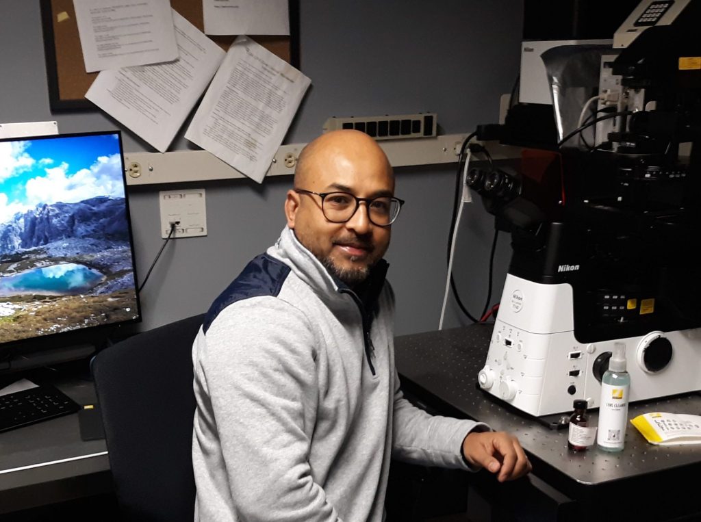
WIDE-FIELD MICROSCOPY
Nikon Ti 2000 wide-field microscope
Capabilities: This microscope is suited for live cell imaging (4D) and fixed cells (3D) imaging. This microscope produces high quality wide-field images that may be improved by deconvolution.
• Special features and accessories: Definite focus, CO2/heating control stage insert, environmental chamber for live imaging.
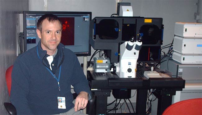
SUPER RESOLUTION MICROSCOPY
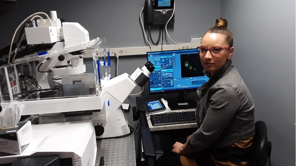
Carl Zeiss ELYRA microscope (PALM/SIM)
Capabilities: ELYRA PS.1 is a SR microscope system for Photoactivated Localization Microscopy (3D PALM) and Structured Illumination Microscopy (SIM), in other words two instruments in one. Depth of focus for 3D PALM based on double-phase ramp is 1.4 um in three dimensions. Expected resolution in XYZ for PALM is 20 nm/ 20 nm / 60 nm. Expected resolution for SIM is 120/120/350 nm. The regular confocal microscopes have a XYZ resolution of 240 nm/ 240 nm/ 700 nm.
• Special features and accessories: Double-phase ramp for 3D PALM, Definite focus, Environmental chamber.
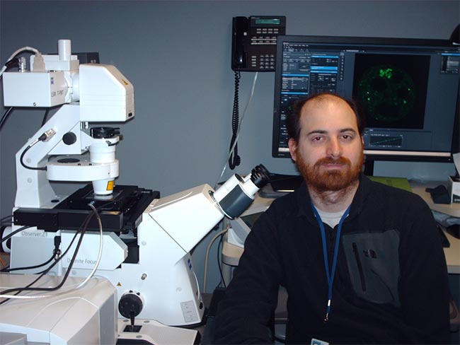
CONFOCAL MICROSCOPY
Carl Zeiss LSM780 Microscope
• Capabilities: Carl Zeiss LSM780 with 34 spectral detectors including high-sensitive GaASP is a point scanning confocal microscope that generates optical sections within 3D specimens. It can collect images in five colors (blue, green, orange, red and far red) and it is especially well suited for multicolor imaging, FRET, photoactivation with 405 nm laser line, and biophysical assays such as FRAP and Number and Brightness.
• Special features and accessories: Definite focus, CO2/heating control stage insert.
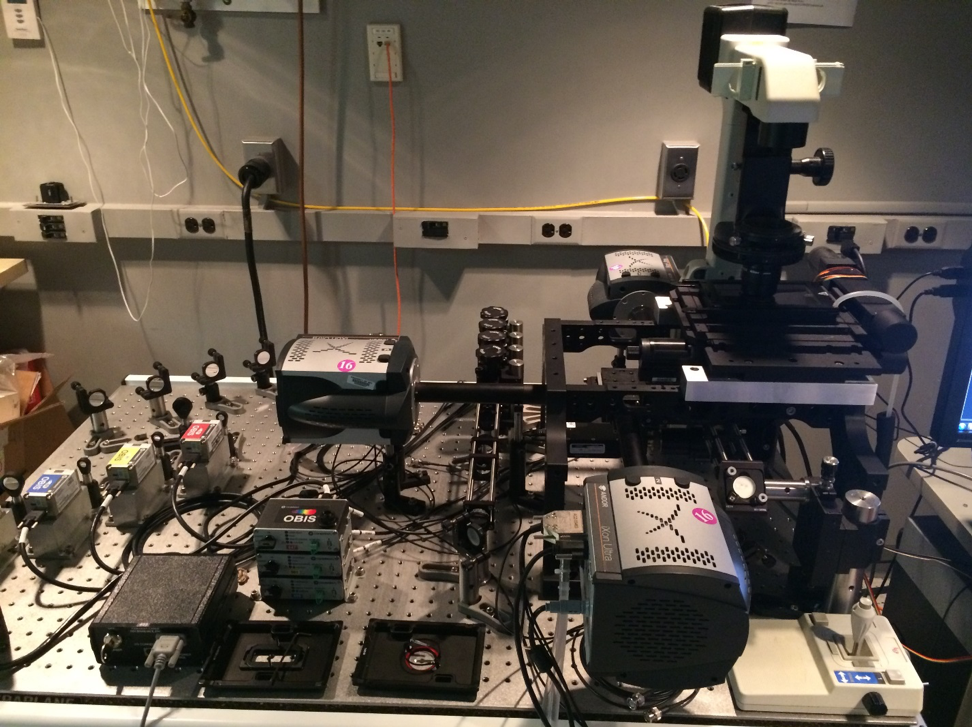
Highly Inclined Laminated Optical Sheet (HILO) MICROSCOPY
Custom-Built Single Molecule Tracking (SMT) Microscope
Capabilities: HILO (highly inclined laminated optical sheet) technique substantially reduces out-of-focus light, increases signal-to noise ratio and allows observation of low light signal; thus, it allows observation and tracking of individual molecules (SMT). This microscope is suited for tracking of single molecules in two color channels using a third channel for a counter-stain. This system also may be used for the fast live imaging. Z-sectioning is currently not available on this system.
• Accessories: OKO lab stage insert with temperature and CO2 control.
CONFOCAL MICROSCOPY
Carl Zeiss Airyscan LSM880 confocal microscope
• Capabilities: Airyscan Zeiss LSM880 confocal microscope is capable of the conventional confocal imaging (XYZ resolution 240 nm / 240 nm / 700 nm). In addition, it is equipped with Airyscan module. This module improves resolution in XYZ beyond the diffraction limit for the live cultures and small animals (zebrafish), providing resolution of 120 nm / 120 nm / 380 nm, or 145 nm / 165 nm / 450 nm in Fast Airyscan mode. 730 nm laser line is available.
• Special features and accessories: Definite focus, CO2/heating control stage insert, environmental chamber for live imaging.
LATTICE LIGHT SHEET (LLS) MICROSCOPY
LLS (Lattice Light sheet) Microscope
• Capabilities: Simultaneous two-color imaging in 3D and 4D producing large volumes of data; long-time imaging without photodamage and phototoxicity with high resolution (XYZ is 230 x 230 x 370 nm). The system generates an optical lattice to convert laser beam into an ultra-thin 0.4 um light sheet consisting of a parallel array of the light beams. In comparison to other imaging methods, the lattice light sheet technique (1) substantially increases a signal to noise ratio and reduces the light exposure, (2) allows ultra-thin optical sectioning (3) ensures extremely low phototoxicity, (4) substantially increases a signal to noise ratio, (5) allows very fast time lapse imaging on the scale of milliseconds (6) increases resolution. Currently this system is suitable only to cell cultures and thin tissue sections, but its capabilities will be eventually expanded to small animals or organs by equipping it with adaptive optics.
• Accessories: OKO lab stage insert with temperature and CO2 control.
MINFLUX (MINimising Fluorescence FUXes) nanoscopy
MINFLUX (MINimising Fluorescence FUXes) nanoscope
• Capabilities: Simultaneous two-color imaging in 3D and 4D with nanoscopic resolution (up to 3 nm in XYZ). This system may be used (a) for molecular mapping of the intracellular structures such as organelles, high molecular complexes, (b) nanometer-scale ultrafast microsecond dynamics of individual molecules associated with intracellular targets such as DNA, transcription factories, nuclear pores, (c) fast micrometer-scale millisecond dynamics of highly mobile (diffusing, gliding, translocating) molecules. The system provides long-time imaging without photodamage and photobleaching. This microscope also is capable of confocal and wide-field epifluorescence imaging.
MINFLUX technology is based on excitation of the fluorescent molecules with doughnut-shaped beam, and photon-counting. Exact position of the molecule is detected by a minimum of fluorescence; thus, it requires low photon count and imaging is fast and non-photodamaging. Resolution is not dependent on wavelength and molecular tilt. MINFLUX technology is based on excitation of the fluorescent molecules with doughnut-shaped beam, and photon-counting. Exact position of the molecule is detected by a minimum of fluorescence; thus, it requires low photon count and imaging is fast and non-photodamaging. Resolution is not dependent on wavelength and molecular tilt. • Accessories: OKO lab stage insert with temperature and CO2 control.