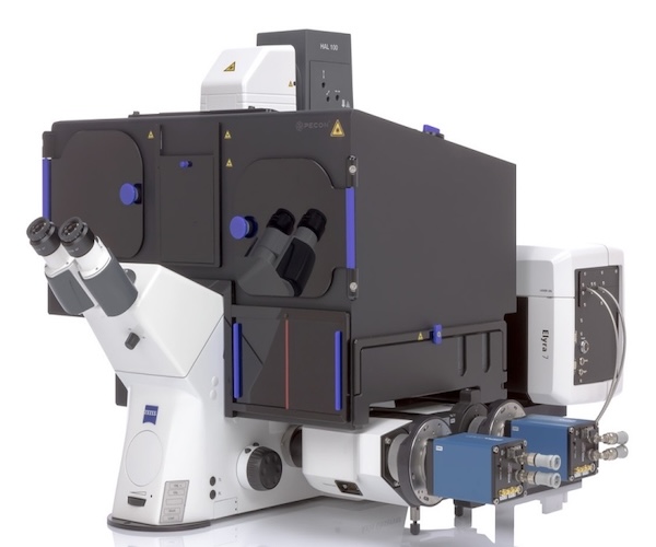
ZEISS Elyra 7 with Lattice SIM²
Type: Wide-field structured illumination microscope
Capabilities:
- Super resolution 2D and 3D imaging of live or fixed cells
- Apotome SIM: 170 nm lateral, 450 nm axial
- Apotome SIM2: 140 nm lateral, 300 nm axial
- Lattice SIM: 120 nm lateral, 300 nm axial
- Lattice SIM2: 60 nm lateral, 200 nm axial
- Simultaneous 4 color acquisition (2 colors/camera)
Laser Lines:
- 405 nm, 458 nm, 561 nm, 642 nm
Objectives (Apotome):
- 10x, dry, 0.30 NA, 5.2 mm
- 20x, dry, 0.80 NA, 0.55 mm
- 40x, water, 1.2 NA, 0.28 mm
- 40x, oil, 1.4 NA, 0.13 mm, DIC
Objectives (SIM):
- 63x, oil, 1.4 NA, 0.19 mm, DIC
- 63x, oil, 1.46 NA, 0.1 mm
Detectors:
- 2 pco.edge sCMOS cameras
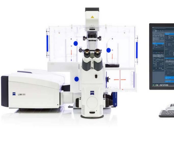
Zeiss LSM 880 Airyscan
Type: Laser scanning confocal microscope
Capabilities:
- Super-resolution confocal imaging – resolution of 140nm, xy and 350nm, z in fixed and live 2D/3D cells
- Improved sensitivity imaging (Improved signal to noise) – 4x to 8x more sensitive than Zeiss LSM 780 in fixed and live 2D/3D cells
- Faster imaging – 27 frames per second at 480 x 480 pixels in fixed and live cells
- Fluorescent Correlation Spectroscopy (FCS)
Laser Lines:
- 405, 458, 488, 514, 561, 594, 633 nm
Objectives:
- 10x, dry, 0.45NA, 2.0 mm
- 20x, dry, 0.80NA, 0.55 mm
- 40x, water, 1.2NA, 0.28 mm
- 40x, oil, 1.3NA, 0.21 mm
- 63x, oil, 1.4NA, 0.19 mm
- 63x, multi-immersion, 1.2NA, 0.49 mm
- 20x, dry, 0.80NA, 0.55 mm
- 40x, water, 1.2NA, 0.28 mm
- 40x, oil, 1.3NA, 0.21 mm
- 63x, oil, 1.4NA, 0.19 mm
- 63x, multi-immersion, 1.2NA, 0.49 mm
Detectors:
- 2) Multialkali photomultiplier tube (MA-PMT)
- Spectral Detector – 32 Channel Gallium Arsenide Phosphide Photomultiplier Tube (GaAsP PMT)
- Airyscan detector – array of 32 individual GaAsP-PMT elements that act as a pinhole size of 0.2 Airy units (AU)
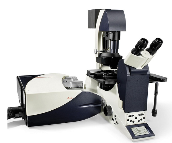
Leica SP8 with Digital Lightsheet
Type: Laser scanning confocal with digital lightsheet microscope
Capabilities:
- 2D / 3D imaging of fixed or live fluorescence-labeled samples
- Imaging of interactions and dynamics of fluorescence labeled molecules
- Digital light sheet imaging of cleared large 3D samples both fixed and live
- Spectral Imaging
- Forster Resonance Energy Transfer (FRET)
- Fluorescence Recovery After Photobleaching (FRAP)
- Laser Lines
Laser Lines:
- 405, 488, 552, 638 nm
Objectives:
- 1.6x, dry, 0.05NA*
- 2.5x, dry, 0.07NA, 9.2 mm*
- 5x, dry, 0.15NA, 12 mm
- 5x, multi-immersion, 0.15NA#
- 10x, dry, 0.4NA, 2.2 mm*
- 10x, water, 0.3NA, 3.6 mm#
- 20x, multi-immersion, 0.75NA, 0.67 mm
- 25x, water, 0.95NA, 2.5 mm#
- 40x, dry, 0.85NA, 0.21 mm
- 63x, water, 1.2NA, 0.30 mm,
- 63x, oil, 1.4NA, 0.14 mm
- 100x, oil, 1.4NA, 0.13 mm
- *Illumination objectives for Digital Lightsheet
- #Detection objectives for Digital Lightsheet
- TwinFlect Mirrors for Digital Lightsheet
- 7.8 mm width; Sample Immersion Liquid – Glycerol, Water; Illumination Obj. – 1.6x, dry; Detection Obj. 5x, multi-immersion or 10x, water
- 5 mm width; Sample Immersion Liquid -Water; Illumination Obj. – 2.5x, dry; Detection Obj. 10x, water or 25x, water
- 2.5 mm width; Sample Immersion Liquid -Water; Illumination Obj. – 2.5x, dry or 10x, dry ; Detection Obj. 10x, water or 25x, water
Detectors:
- (1) Photomultiplier tube
- (2) Hybrid detectors (HyD)
Instructions:
- Turn On/Turn Off (PDF, 647 KB)
- Basic Image Acquisition (PDF, 7MB)
- Analysis (PDF, 2MB)
- How to Change Bit Depth in LASX Software (PDF, 511KB)
- Imaging (PDF, 1MB)
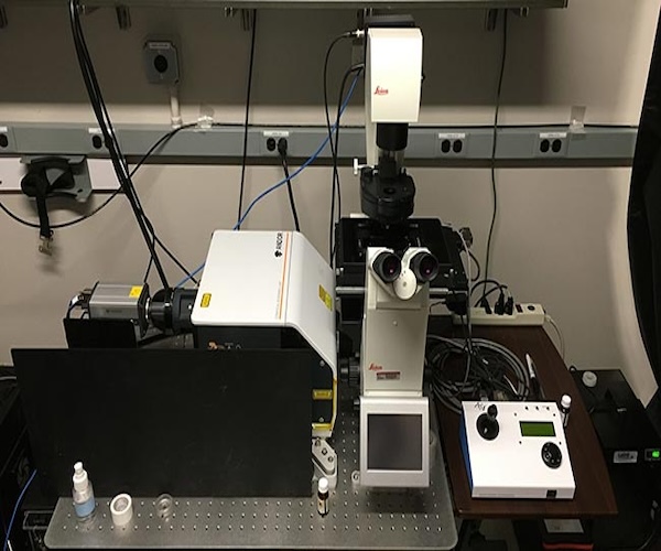
Andor spinning disk confocal on Leica DMi8 Base
Type: Spinning disk confocal
Capabilities:
- 2D / 3D imaging of fixed or live fluorescence-labeled samples
- Imaging of interactions and dynamics of fluorescence labeled molecules
- Large area montages
Laser Lines:
- 405, 445, 488, 514, 561, 594, 640, 730 nm
Objectives:
- 1.6x, dry, 0.05NA
- 5x, dry, 0.15NA, 12 mm
- 10x, dry, 0.4NA, 2.2 mm
- 20x, dry, 0.75NA, 0.62 mm
- 63x, water, 1.2NA, 0.30 mm,
- 63x, oil, 1.4NA, 0.14 mm
Detectors:
- Andor Xyla sCMOS camera
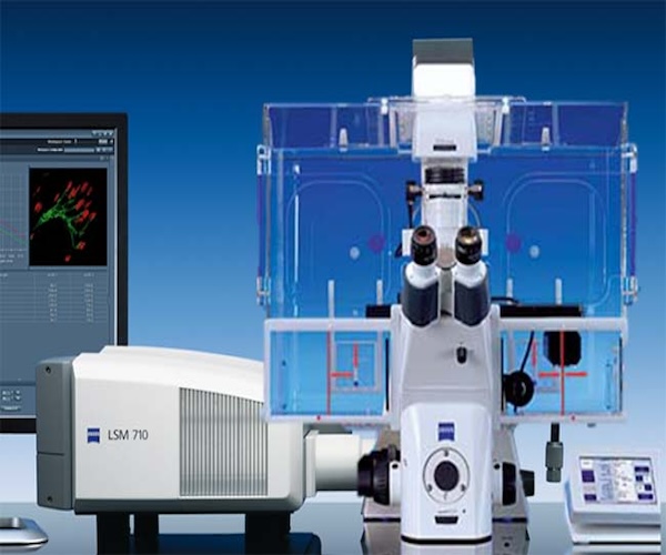
Zeiss LSM710
Type: Laser scanning confocal microscope
Capabilities:
- 2D / 3D imaging of fixed or live fluorescence-labeled samples
- Imaging of interactions and dynamics of fluorescence labeled molecules
Laser Lines:
- 405, 458, 488, 514, 561, 594, 633, 690 – 1000 nm (pulsed)
Objectives:
- 5X, dry, 0.15 NA, 13.6 mm
- 10X, dry, 0.45 NA, 2.00 mm
- 10X, water, 0.45 NA, 1.8 mm
- 20X, dry, 0.4 NA, 7.9 mm
- 40X, water, 1.1 NA, 0.62 mm 63X, oil, 1.4 NA, 0.19 mm
Detectors:
- Photomultiplier tubes
- Spectral detector
Instructions:
- Turn On Instructions (PDF, 355 KB)
- OMAL Server Connection on LSM710 (PDF, 468 KB)
- RTC Error Message at Start Up (PDF, 325 KB)
- LSM710 DIC Tutorial >(PDF, 2 MB)
- LSM710 Live Cell Turn On (PDF, 858 KB)
- Avoiding Focus Drift (PDF, 1 KB)
- Bleaching on the LSM710 (PDF, 462 KB)
- FRAP on LSM710 and LSM780> (PDF, 744 KB)
- Single Position Time Series on the LSM710 (PDF, 394 KB)
- Multi Position Time Series on the LSM710 (PDF, 498 KB)
- Autofocus on the LSM710 (PDF, 282 KB)
- Autofocus Tiling on the LSM710 (PDF, 1 MB)
- Lambda Mode (PDF, 424 KB)
- Tiling on Zeiss Microscopes (PDF, 460 KB)
- 710 Troubleshooting (PDF, 598 KB)
- Post Processing of Images LSM710 (PDF, 791 KB)
Zeiss LSM780
Type: Laser scanning confocal microscope
Capabilities:
- 2D / 3D imaging of fixed or live fluorescence-labeled samples
- Imaging of interactions and dynamics of fluorescence labeled molecules
- Live animal imaging
Laser Lines:
- 405, 458, 488, 514, 561, 633, 690 – 1000 nm (pulsed)
Objectives:
- 10X, dry, 0.3 NA, 5.1 mm
- 20X, dry, 0.8 NA, 0.55 mm
- 20X multi-immersion
- 20X water (dipping lens, not on turret), 1.0NA, 0.19 mm
- 40X, water, 1.1 NA, 0.28 mm
- 40X, oil, 1.3 NA, 0.2 mm
- 63X, oil, 1.4 NA, 0.19 mm
Detectors:
- Photomultiplier tubes
- Spectral detector
- Non-descanned detectors (for use with 2P laser)
- Fluorescence life-time detectors
Instructions:
- LSM780 Turn On/Turn Off Instructions (PDF, 436 KB)
- LSM780 DIC Tutorial (PDF, 850 KB)
- Live Cell Turn On/Turn Off Instructions (PDF, 1.0 MB)
- LSM780 Lambda Mode (PDF, 747 KB)
- Avoiding Focus Drift (PDF, 5 KB)
- Single Position Definite Focus on the LSM780 (PDF, 529 KB)
- Multi Position Definite Focus on the LSM780 (PDF, 823 KB)
- LSM780 Multi Time with Definite Focus (PDF, 1 MB)
- FLIM (PDF, 2.3 MB)
- FRAP on LSM710 and LSM780 (PDF, 744 KB)
- Convex Hull Tile Scanning on the LSM780 (PDF, 102 KB)
- OMAL Server Connection on LSM780 (PDF, 556 KB)
- Post Processing of Images on LSM780 (PDF, 713 KB)

Nikon STORM
Type: Widefield super-resolution microscope for fixed cells only
Capabilities:
- Super-resolution by Point Localization Microscopy – resolution 20nm in xy and 50nm in z.
- Recommended modality is dSTORM with CF-647 dye (improved version of Alexa Fluor 647 dye).
Laser Lines:
- 405, 488, 561, 647 nm
Objectives:
- 100x oil, 1.49 NA, 0.13 mm WD
- 100x silicon oil, 1.35 NA, 0.3 mm WD
Cameras:
- Andor iXON DU-897U
Nikon SIM
Type: Wide-field super-resolution microscope for fixed cells only
Capabilities:
- N-SIM: Super-resolution Structural Illumination Microscopy (SIM) – resolution of 110nm in xy and 275nm in z. Compatible with conventional immunofluorescent staining.
- “External-phase” trans-illumination imaging (on side eye-port camera), which is useful for correlative light/electron microscopy. None of the high NA objectives on this microscope are compatible with regular phase imaging.
Laser Lines:
- 405, 488, 561, 640 nm
Objectives:
- 10x, dry, 0.3 NA, 16 mm WD
- 60x water, 1.27 NA, 0.17 mm WD
- 100x oil, 1.49 NA, 0.13 mm WD
Cameras:
- Andor iXON DU-897E for SIM
- Nikon DS-QiMC for external phase microscopy
Nikon Multifunctional Microscope (MFM)
Type: Widefield
Capabilities:
- 2D / 3D imaging of fixed or live fluorescence-labeled samples
- High content analysis
- TIRF
- Polarization imaging
- Simultaneous 2-color imaging
- Single molecule imaging
Excitation lines:
- (LED illuminator)
- 395, 440, 470, 508, 555, 640 nm
Objectives:
- 4X, dry, 0.13 NA, 16.5 mm
- 20x, dry, 0.75 NA, 1.00 mm
- 60X, oil, 1.40 NA, 0.13 mm
- 100X, oil, 1.45 NA, 0.13 mm
Detectors:
- Andor NeoZyla sCMOS camera
- Andor iXon Ultra EMCCD camera
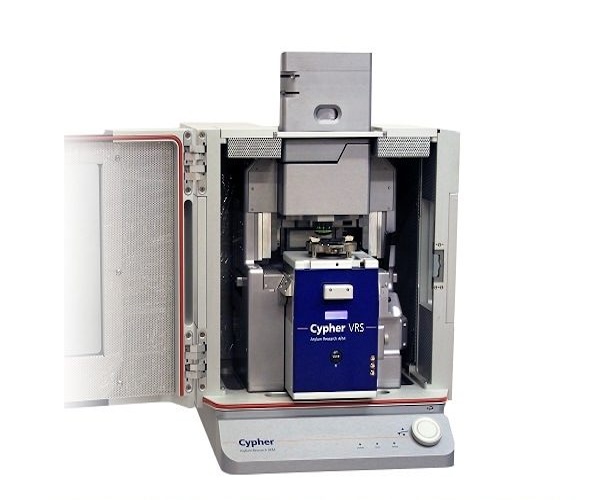
Asylum Research Cypher-VRS
Type: Atomic force microscope
Capabilities:
- Video-rate scanning for molecular dynamics imaging
- Contact and tapping mode imaging in air and liquid environments
- Force spectroscopy
- Mechanical mapping
- Temperature control
- Perfusion chamber
- Semi-automated probe alignment and calibration
- Built-in analysis tools
Objectives:
- 20X reflective objective for cantilever setup and sample locating
Sample Formats:
- Standard imaging: 10 mm diameter substrate
- Video-rate imaging: 3 mm diameter substrate
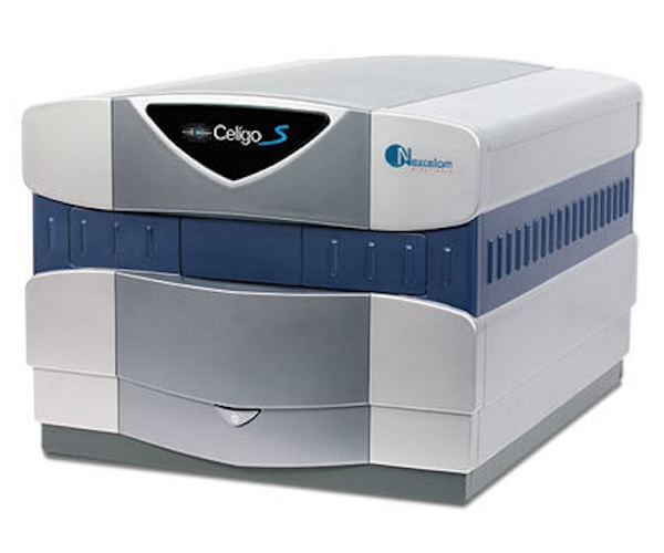
Celigo
Type: Plate reading microscope
Capabilities:
- Built in analysis tools
- Fast, widefield imaging of entire well for multiwell plate
- Built in protocols for live/dead counting, growth curves, spheroid analysis etc.
Excitation/Emission Filters:
- Blue 377/477
- Green 483/536
- Red 531/629
- Far Red 632/692
- Bright-field
Objectives:
- 1 objective optimized for entire well imaging & built in analysis features
Sample Formats:
- Works best with supported/recommended plates
- Microwell plates: 6, 12, 24, 48, 96, 384, 1536 wells
- Slides
- 10 cm Petri Dish
- T25 Flasks
Instructions:
- Nexcelom Reagents (Links)
- Recommended and Supported Plates (PDF, 170 KB)