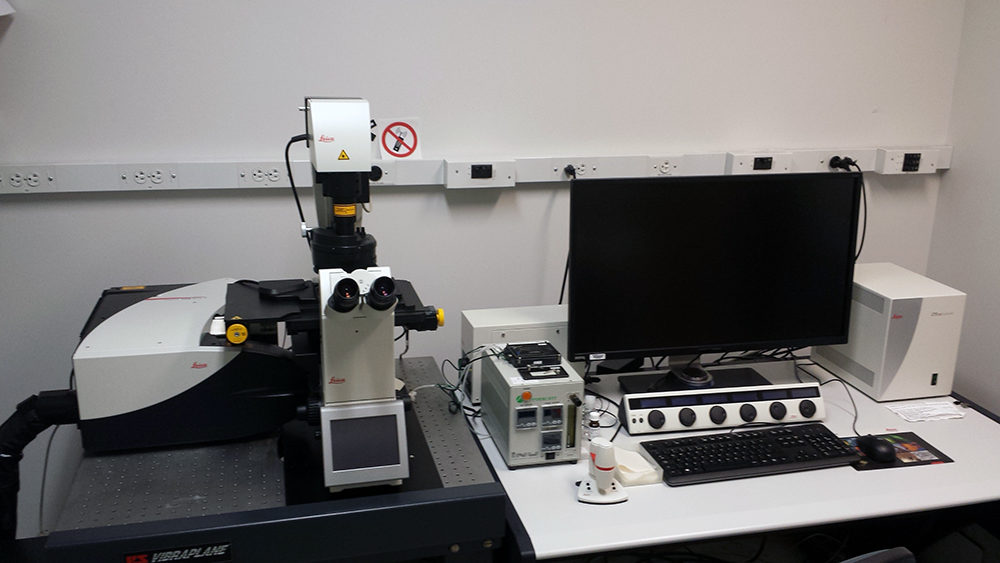
Leica SP8 LSCM with white light laser
The SP8 LIGHTNING confocal microscope allows you to make proper and detailed observations of fast biological processes. Your experimental work will have the benefit of super-resolution, high-speed imaging, and the capability to image multiple fluorescent markers simultaneously.
Thanks to the exclusive detection concept of LIGHTNING, you can trace the dynamics of multiple molecules, even those expressed at low levels, simultaneously over long recording times in living specimens.
The SP8 LIGHTNING confocal microscope offers you the following The SP8 LIGHTNING confocal microscope offers you the following performance advantages
• truly simultaneous multicolor imaging in super-resolution down to 120 nm
• live specimen imaging thanks to fast acquisition rates
• sample protection thanks to low phototoxicity
The SP8 LIGHTNING confocal microscope opens up unique experimental options to inspire your research!


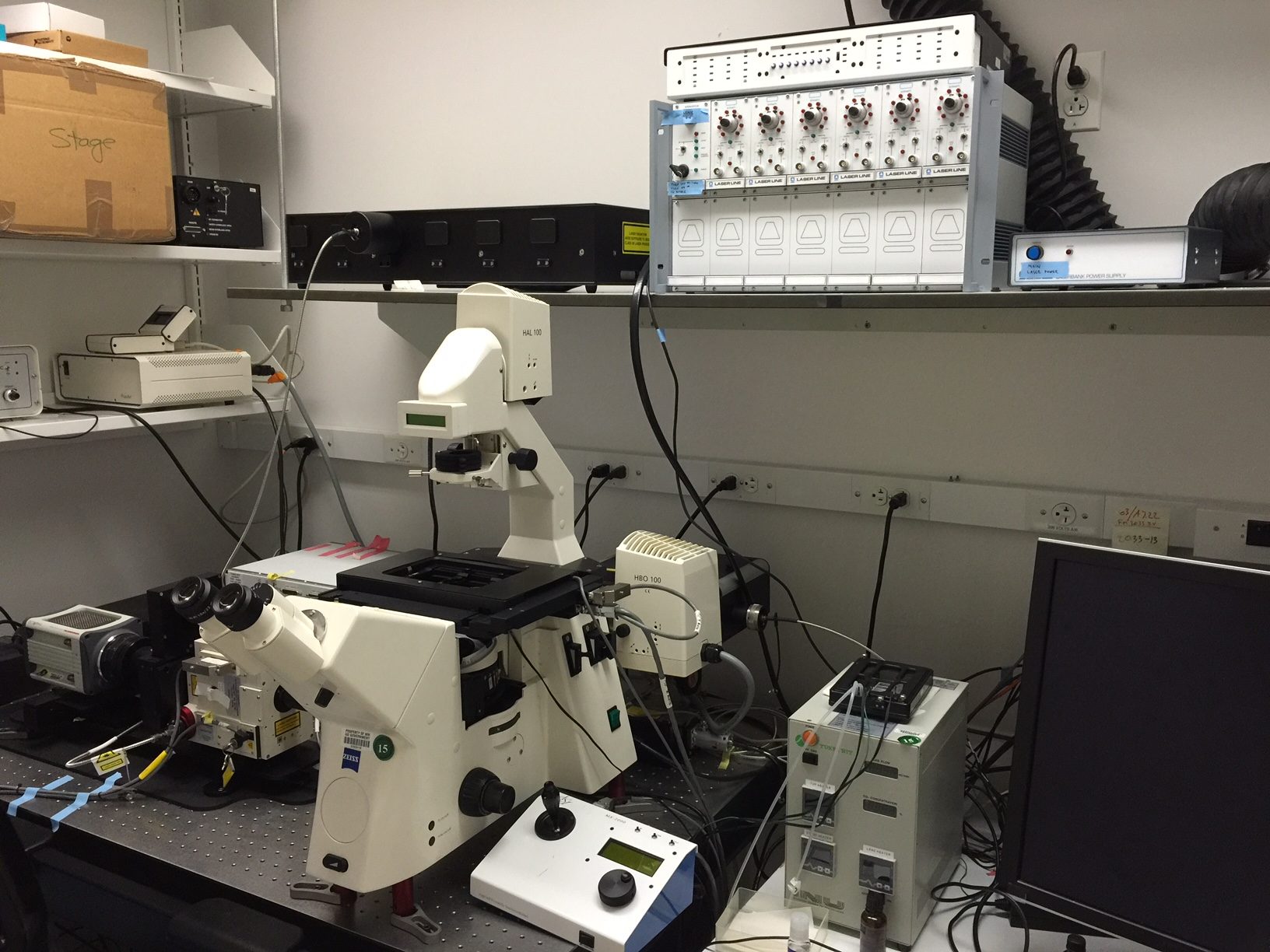
Spinning disk confocal
In conventional widefield optical microscopy the specimen is bathed in the light used to excite fluorophores. The fluorescence emitted by the specimen outside the focal plane of the objective interferes with the resolution of in focus features. As the sample increases in thickness, the ability to capture fine detail above out-of-focus signal becomes increasingly challenging. The confocal principle rejects out-of-focus light through the use of a pinhole, with the added benefit of an improvement in lateral and axial resolution.
LEARN MORENikon TIRF
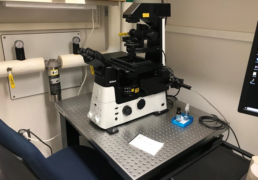
Various mechanisms are often employed in fluorescence microscopy applications to restrict the excitation and detection of fluorophores to a thin region of the specimen. Elimination of background fluorescence from outside the focal plane can dramatically improve the signal-to-noise ratio, and consequently, the spatial resolution of the features or events of interest.
Total internal reflection fluorescence microscopy (TIRFM) exploits the unique properties of an induced evanescent wave or field in a limited specimen region immediately adjacent to the interface between two media having different refractive indices. In practice, the most commonly utilized interface in the application of TIRFM is the contact area between a specimen and a glass coverslip or tissue culture container.

