Software
Several software packages are available to users
Arivis Vision 4D
Arivis Vision4D is a modular software for working with multi-channel 2D, 3D and 4D images of almost unlimited size independent of available RAM.
LEARN MORE
Arivis InViewR VR (Virtual Reality)
arivis InViewR sets new standards for analyzing Life Science research images. Freed from being tethered to a mouse like on a desktop computer and with depth perception equivalent to the real word.
LEARN MORE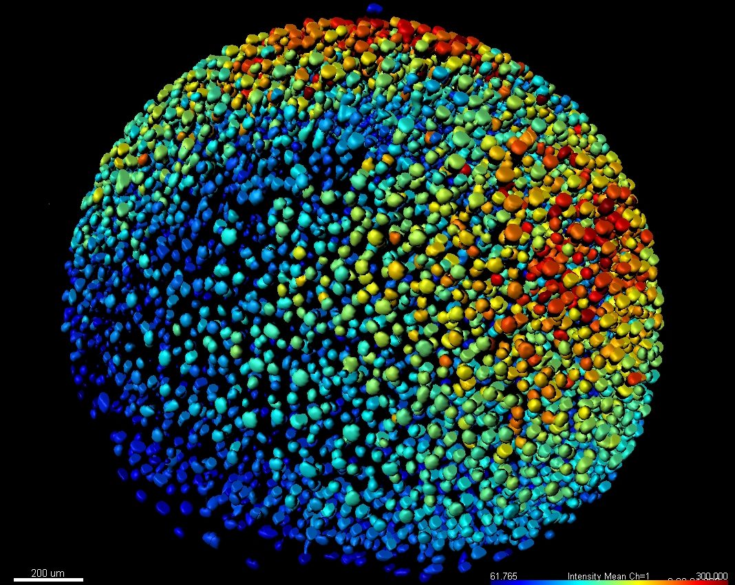
Imaris
The world’s leading Interactive Microscopy Image Analysis software company, actively shaping the way microscopic images are processed through constant innovation and a clear focus on 3D and 4D imaging.
LEARN MORE
Python
Python is a high-level, general-purpose programming language. Its design philosophy emphasizes code readability with the use of significant indentation.
LEARN MORE
Fiji Image J
Fiji is an image processing package—a “batteries-included” distribution of ImageJ, bundling a lot of plugins which facilitate scientific image analysis.
LEARN MORE
Zeiss Zen
Arivis Vision4D is a modular software for working with multi-channel 2D, 3D and 4D images of almost unlimited size independent of available RAM.
LEARN MORE
Nikon Elements
NIS-Elements Viewer is a free standalone program to view image files and datasets. It offers the same powerful view and image selection modes as the NIS-Elements core packages.
LEARN MORE
Leica LAS-X
LAS X microcope software for all Leica microscopes: It integrates confocal, widefield, stereo, super-resolution, and light-sheet instruments.
LEARN MORE
R for image analysis
R is a free software environment for statistical computing and graphics. It compiles and runs on a wide variety of UNIX platforms, Windows and MacOS.
LEARN MORETechniques
Techniques used by the Core include:
FRAP(Fluorescence Recovery After Photobleaching)
Is a method for determining the kinetics of diffusion through tissue or cells. It is capable of quantifying the two dimensional lateral diffusion of a molecularly thin film containing fluorescently labeled probes, or to examine single cells.
FRET (Fluorescence Resonance Energy Transfer)
FRET is a technique to detect protein interaction and the formation of complexes between different proteins tagged with fluorescence markers. It also can be used to detect enzymatic activity by special reporters (FRET biosensors).
FLIM (Fluorescence-lifetime imaging microscopy)
Is an imaging technique for producing an image based on the differences in the exponential decay rate of the fluorescence from a fluorescent sample.
Photoactivated localization microscopy (PALM)
Photoactivated localization microscopy (PALM) is a Super Resolution (SR) technique based on the photoactivation of photoconvertable fluorescent molecules fused to the protein of interest.
Multi-photon Imaging
Two-photon excitation microscopy is a fluorescence imaging technique that allows imaging of living tissue up to about one millimeter in depth. It differs from traditional fluorescence microscopy, in which the excitation wavelength is shorter than the emission wavelength, as the wavelengths of the two exciting photons are longer than the wavelength of the resulting emitted light.
Structured illumination (SIM)
Structured illumination (SIM) is a Super Resolution (SR) technique based on imaging of a set of images from the same focal plane with a shifting grid pattern. Information is extracted from the raw data to produce a reconstructed image having a lateral resolution of 100 nm (approximately twice that of conventional confocals and wide-field instruments) and an axial resolution ranging between 150 and 300 nanometers.
Light Sheet FluorescenceMicroscopy
Light sheet fluorescence microscopy is a fluorescence microscopy technique with an intermediate-to-high optical resolution, but good optical sectioning capabilities and high speed. It is used for imaging live, fixed or cleared biological samples
Confocal Microscopy
For imaging fluorescently labeled specimens and permitting accurate optical sectioning for volumetric studies, such as large extended field of view tile imaging of tumor samples.
Photoswitching
Of specialized fluorescent proteins to monitor the dynamics of sub-cellular structural components by live cell Airyscan super-resolution microscopy.
